Orthopädie am Rhy

Basel, Rheinfelden, Liestal

What are facet joints?

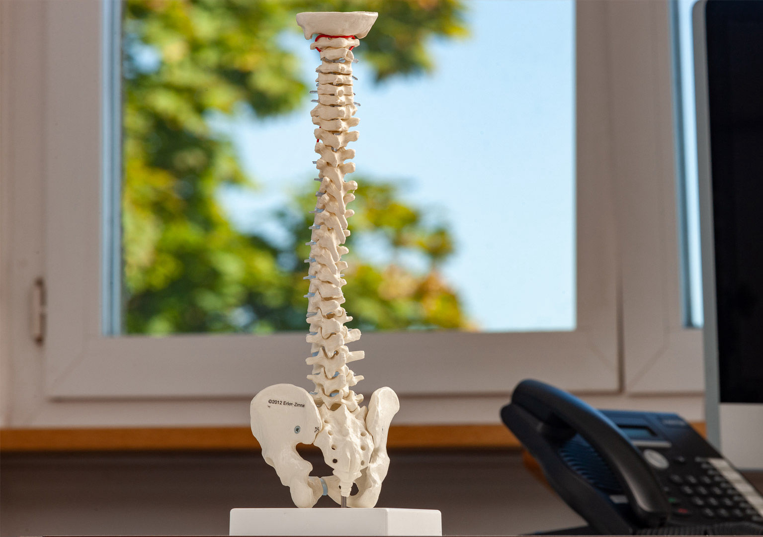
The facet joints are small paired joints at the top and bottom of each of the vertebral arches which enclose the spinal canal. Together with the intervertebral discs between the individual vertebral bodies, they enable segmental movements of each individual vertebra in the lumbar spine.

Like all joints, spinal facet joints have a cartilage surface and a connective tissue joint capsule. The joint capsules of the lumbar vertebrae are covered by a firm, yellowish connective tissue inner fascia, the ligamentum flavum (= yellow band), which runs alongside the spinal canal. Between two facet joints of the lower lumbar spine, the sciatica nerve roots run from the leg through the spinal canal to the brain. 
The typical symptoms of facet syndrome are:
• acute, chronic or episodic back pain
• stronger pain during exertion and movement
• sometimes radiating pain to the buttocks, hips and legs (spondylogenic)
• painfully restricted movement in the lower back
• painfully cramped deep back muscles (Musculi Multifidi)
• it is not uncommon for there to also be radiating pain along the sciatic nerve (pseudoradicular) 

Facet syndrome can be diagnosed very well as the cause of deep back pain. Magnetic resonance imaging (MRI) in particular, and rarely also computed tomography CT, are used to identify and assess arthritis or activated arthrosis of the lumbar vertebrae and sacral joints, lumbar spine and SI joint, in the case of persistent lower back pain and deep hip pain. The vertebral joints of the lumbar spine can only be palpated indirectly. In a physical exam, special tests can provide good information. If there is justified suspicion by an osteoarthritis and rheumatism specialist for the diagnosis of "activated facet syndrome" or "ISJ syndrome", a special magnetic resonance MRI is mostly commonly used today. For further confirmation of the diagnosis and also for the treatment, a diagnostic and therapeutic test infiltration by means of targeted injection of a drug that is only effective there can be carried out under image control with modern ultrasound or image converter devices suitable for this purpose.

Painkillers often only relieve symptoms temporarily. Painkillers are usually unable to adequately cure the sometimes extremely painful swelling of the vertebral joints caused by osteoarthritis, arthritis and myofibrosis. Recurring facet syndromes often result in significant permanent restrictions of movement due to scarring of the vertebral joints and the deep fasciae and atrophy of the muscles of the lower back. The goal is to break this vicious circle. Very effective therapeutic interventions are available today to treat facet joint and/or SI joint syndrome. The prerequisite, however, is that a facet syndrome must be confirmed as the cause of pain and located with certainty. Image-controlled facet infiltrations of various drugs, such as cortisone or biostimulative substances, into the facet joints and around the sciatic nerve roots with its ganglia of the deep fascia. Other methods include neurostimulation and neuromodulation, electrophysiologically controlled neurolysis of the medial branches of the lumbar and sacrum joints. A controlled and adapted personalised reconditioning according to the above-mentioned procedures is used to restore as much agile and resilient mobility as possible. During the consultation, the indications and therapeutic interventions are of course explained in detail! They can almost always be carried out on an outpatient basis.

Your spine specialists in our practice

Dr Moritz Dau

Specialist FMH, Sports medicine, SEMS
Interventional pain management SSIPM
Ultrasound on the musculoskeletal system SGUM 
Manual medicine/Chirotherapy SAMM
Entire musculoskeletal system & spine


Jaroslaw Boronzcyk

Specialist in Orthopaedics


© 2024 Copyright Orthopädie am Rhy

LOCATIONS